Achieve clearer margins, accurate contacts, and more predictable restorations.
Intraoral scanning (IOS) has transformed digital dentistry — but even the best scanners rely on strong fundamentals. Small errors in technique can lead to unclear margins, incomplete contacts, and inaccurate occlusion. The good news? Most scan issues can be prevented with simple, consistent habits. Many new IOS systems have a Digital Denture module button to help you with edentulous areas
Here are SCD’s essential tips and troubleshooting steps to help you capture accurate scans every time.
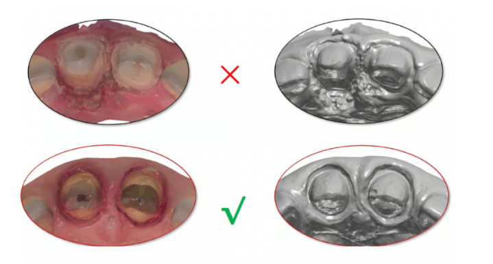
1. Capture Clear, Dry, Well-Defined Margins
The most common cause of remakes is unclear or incomplete margins.
Tips
- Thoroughly clean and air-dry the prep before scanning
- Ensure the scanner lens is clean and dry
- Maintain the correct distance between the scanner tip and the preparation
- Move slowly and deliberately – rushing increases stitching errors
Clear, uninterrupted margins provide the foundation for accurate restorations.
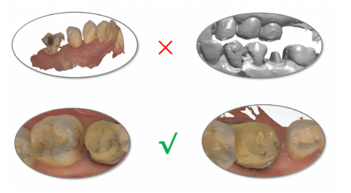
2. Scan Approximal Contacts Completely
Missing contact areas lead to poor fit and inaccurate proximal contours.
Tips
- Perform a full arch scan with the scanbody in place
- Then remove the scanbody and capture approximal areas separately
- Check for stitching gaps or voids before sending the file
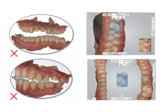
3. Scanning Patients with Dentures
Clasps and denture surfaces are often shiny, causing incomplete data capture.
Tips
- Apply a scan spray or matting agent to the denture – see if your supplier stocks this
- Complete an additional scan of the denture alone, including the fitting surface
This ensures accurate reference for occlusion and clasp contouring.
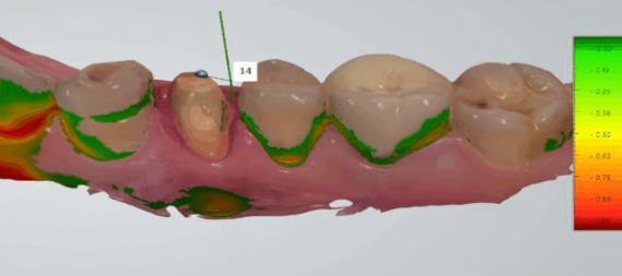
4. Review Undercuts Before Sending
Undercuts can distort emergence profiles or cause path-of-insertion issues.
Tips
- Use the scanner’s undercut measurement tool
- Re-prepare and re-scan any identified areas
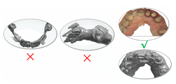
5. Ensure Full Margin Coverage
During your review phase, verify the entire circumference is fully captured.
Tips
- No holes or gaps
- Smooth, continuous margin line
- No shadowing from gingiva or saliva
If any part is unclear → re-scan that area immediately.
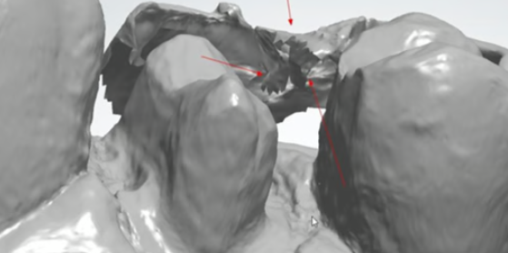
6. Confirm Approximal Contacts
Incomplete contacts are a frequent source of inaccuracies.
Tips
- Missing tooth structure
- Gaps, folds, or holes in approximal anatomy
- Proper stitching between buccal and lingual passes
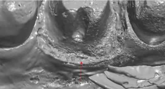
7. Pontic Sites Must Be Fully Captured
Pontic areas often get overlooked in fast scanning.
Tips
- Slow down and ensure full 360° capture of the pontic site, including ridge contour and soft tissue.
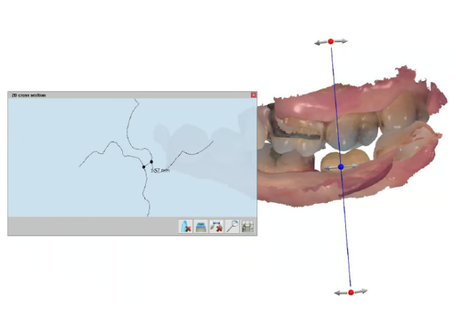
8. Check Occlusal Clearance
Use the 2D cross-section tool to verify there is adequate space.
Tips
- Confirm clearance on both functional and non-functional cusps
- If insufficient, adjust the prep and re-scan
9. Scan Bite From Both Sides
One-sided bite scans can introduce occlusal discrepancies.
Tips
- Always capture left and right bite scans to improve occlusal accuracy and reduce open-bite errors.
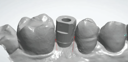
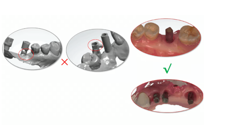
10. Scanbodies Must Be Fully Captured
In implant workflows, the scanbody’s geometry is essential.
Check
- Full 360° capture
- Clear edges and anti-rotational features
- No stitching errors around the cylinder
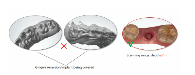
11. Prevent Gingival Recession Around Implants
Delays during scanning can cause tissue collapse or distortion.
Tips
- Scan approximal areas first
- Remove the healing abutment and scan the gingiva immediately
- Move with purpose — don’t pause too long near the site
Scanning range depth should generally not exceed 7 mm.
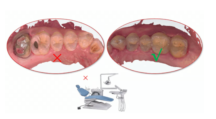
12. Avoid External Light Interference
Strong light sources can create scan noise and shadowing.
Tips
- Reduce overhead lighting
- Avoid direct sunlight
- Maintain a consistent lighting environment
Final Thoughts
Accurate intraoral scans depend on technique just as much as technology. By following these practical tips and taking advice from your scanning company and the rep you have purchased from clear margins, complete contacts, accurate bite capture, and careful implant scanning so you can significantly improve accuracy, reduce remakes, and deliver more predictable outcomes for your patients.